Diagnostics and After Treatment Follow-Up
Several tests are used in the diagnosis of cancer and for monitoring a patient following treatment for appendiceal cancer and peritoneal carcinomatosis. These include:
Biopsy
A biopsy is a test in which tissue from a tumor is removed and visualized under a microscope. A biopsy determines whether a tumor is benign (not cancer) or malignant (cancer). Cancerous tumors are graded from 1 to 4. Low grade tumors are considered less invasive and to grow more slowly, so have a better prognosis. High grade tumors are considered to be faster growing and more invasive, so have a poorer prognosis
- Grade 1 tumors are well-differentiated (resemble normal tissue more closely). These are considered low grade cancers
- Grade 2 tumors are moderately differentiated and are considered intermediate grade tumors
- Grade 3 tumors are poorly differentiated and are considered high grade tumors
- Grade 4 undifferentiated tumors, considered high grade tumors
Tumor Markers
Tumor Markers are substances found in the blood or body fluids that can be created by a cancerous tumor or created by the body in response to a cancerous tumor. Tumor markers can be used to help diagnose cancer and to help monitor the effects of treatments on cancer. Tumor marker tests are never used by themselves to diagnose cancer as sometimes other diseases or conditions other than cancer can cause a tumor markers to be elevated, and other times tumor markers may be absent in the presence of cancer.
Tumor marker are useful, though, in helping to monitor cancer treatment. For instance if a cancer diagnosis has been confirmed by biopsy and cancer tumor markers in the blood are also elevated, a lowering of cancer markers may indicate successful treatment with surgery or a successful response to chemotherapy. In that case, a later rise in tumor marker values after successful treatment may indicate possible recurrence of the cancer. Tumor marker tests that may be used include:
- CEA (Carcinoembryonic antigen) Normal value < 2.5ng/ml, smoker <5.0ng/ml
can be elevated with other non-cancerous diseases such as cirrhosis, inflammatory bowel disease, chronic lung disease, and pancreatitis. Can be positive also in the absence of any disease in a small percentage of the population - CA 125 normal value < 35 kU /ml
can be elevated in ovarian, endometrial, pancreatic, lung, breast, and colon cancers. May also be positive in menstruation, pregnancy, endometriosis, and other conditions. - CA19-9 normal value <40 kU/L
can be elevated in gastrointestinal adenocarcinoma, gastric cancer, colon cancer, and pancreatic cancer
Scans
There are several types of scans that may be used to help visualize cancerous tumors in the body. Scans that may be used in the diagnosis of cancer include:
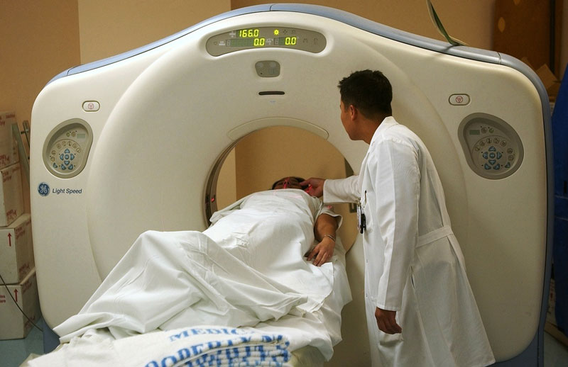
- Computerized tomography scan (CT scan): see the National Cancer Institute’s Computed Tomography (CT): Questions and Answers
- Positron emission tomography scan (PET Scan): A test in which a small amount of radioactive glucose (sugar) is injected into a vein. A computerized scanner then locates areas in the body where the glucose is being used. Since cancer cells use more glucose than normal cells, this test can be used to help locate cancerous tumors in the body. A limitation of PET scans is that other conditions may also cause an increased uptake of glucose (infection, inflammation, trauma, TB) so false positives are common. It is difficult to confirm a cancer diagnosis based on the results of this test alone.
- Octreotide scan: a nuclear medicine scan used to detect carcinoid (neuro-endocrine) tumors
Second-look Laparoscopic Surgery
Secpmd-look laparoscopic surgery is a surgical procedure in which a laparoscope is inserted into a small incision to visualize the interior of the abdomen and to look for the presence of cancerous tumors following cytoreduction (debulking) surgery. This is sometimes done routinely during laparoscopic surgery to reverse a colostomy, and may also be done in other select circumstances. Sometimes visualization may be limited by scar tissue and adhesions from prior surgeries.
Post-operative Follow-up Testing
Following cytoreduction surgery and/or peritoneal chemotherapy and possibly IV chemotherapy for appendix cancer, testing will be done periodically to evaluate for possible cancer recurrence.
- CT scans will usually be done approximately 3 months after surgery initially, then usually every 6 months for the next 3-5 years, followed by yearly scans at the discretion of the surgeon and/or oncologist.
- PET scans may also be done at the discretion of the attending surgeon/oncologist.
- Tumor markers may be evaluated every 3-6 months, again, depending on the preference of the surgeon or oncologist.
- While on chemotherapy and for a period of time following chemotherapy, blood tests will also be done to evaluate the effect of chemotherapy on blood counts.
- A periodic comprehensive physical exam by a physician and evaluation of any symptoms is also extremely important